Exploring the Types of Images Digital Pathology Can Reveal
Digital pathology has revolutionized the way we examine and diagnose diseases at a microscopic level. By converting traditional glass slides into digital images, pathologists can leverage advanced technologies for more accurate and efficient diagnoses. This transformation opens up a world of possibilities, allowing various types of images to be viewed and analyzed. Keep reading to learn about the different kinds of images digital pathology can reveal, highlighting its impact on modern medicine.
Whole Slide Images (WSI)
Whole slide imaging is the cornerstone of digital pathology. This process involves scanning entire glass slides to create high-resolution digital images that can be viewed, navigated, and analyzed on a computer screen. WSIs provide several benefits:
- Comprehensive analysis – Every part of the slide can be examined in detail, ensuring no area is overlooked.
- Remote access – Pathologists can access slides from anywhere, facilitating telepathology and consultations with experts worldwide.
- Enhanced collaboration – Digital slides can be easily shared among colleagues, promoting collaborative diagnostics and research.
Cellular and Tissue Images
Digital pathology allows for the detailed examination of individual cells and tissues, providing insights into their morphology and structure. These images include:
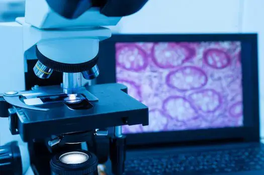
- Hematoxylin and eosin (H&E) stained images – These are the most common types of stained slides used in pathology. H&E staining highlights cellular and tissue structures, aiding in the diagnosis of various conditions, including cancers.
- Immunohistochemistry (IHC) images – IHC uses antibodies to detect specific antigens in cells, providing critical information about protein expression patterns. This is particularly useful in identifying types and origins of tumors.
- Fluorescent stained images – Fluorescent stains help pathologists visualize specific components within cells, such as DNA, RNA, and proteins. These images are invaluable in research settings for studying cellular functions and disease mechanisms.
3D Reconstruction Images
Advancements in digital pathology now allow for the creation of three-dimensional reconstructions from two-dimensional slide images. 3D imaging offers several advantages:
- Spatial understanding – Pathologists can better understand the spatial relationships among different structures within a tissue.
- Detailed analysis – 3D images provide more detailed information about tissue architecture, which can be crucial in understanding complex diseases.
- Improved diagnostics – This technology can enhance the accuracy of diagnoses by revealing features that might be missed in traditional 2D images.
Dynamic and Interactive Images
Digital pathology isn’t limited to static images. Interactive and dynamic imaging techniques include:
- Virtual microscopy – Users can zoom in and out and navigate through digital slides as if using a physical microscope. This enhances the learning experience for medical students and professionals.
- Time-lapse imaging – This involves capturing images at different time points to study dynamic processes, such as cell growth, division, and response to treatments.
- Multiplex imaging – This technique allows for the simultaneous visualization of multiple biomarkers in a single tissue section, providing comprehensive insights into disease pathology.
Computational and AI-Enhanced Images
Artificial intelligence and machine learning are transforming digital pathology by providing computationally enhanced images, which can significantly enhance the efficiency and accuracy of pathology workflows and bioinformatics services. These include:
- Quantitative analysis – AI algorithms can quantify various parameters, such as cell count, tissue density, and biomarker expression levels, providing objective and reproducible data.
- Pattern recognition – AI can detect patterns and anomalies in images that might be subtle or missed by the human eye. This is particularly useful in screening for cancers and other diseases.
- Predictive modeling – AI can analyze image data to predict disease outcomes and responses to treatments, assisting in personalized medicine.
Special Stains and Techniques
In addition to traditional H&E and IHC staining, digital pathology supports a variety of special stains and techniques, such as:
- Periodic acid-Schiff (PAS) staining – Used to detect polysaccharides and mucosubstances in tissues, aiding in the diagnosis of fungal infections and glycogen storage diseases
- Masson’s trichrome staining – Highlights collagen fibers in tissues, useful in evaluating fibrosis and other connective tissue disorders
- Silver staining – Commonly used to visualize nerve fibers, spirochetes, and reticular fibers in tissues
Digital pathology has dramatically expanded the types of images that can be viewed and analyzed, enhancing the capabilities of pathologists and researchers. From whole slide images and detailed cellular structures to advanced 3D reconstructions and AI-enhanced analyses, the potential of digital pathology is vast. This technology not only improves diagnostic accuracy and efficiency but also opens new avenues for research and collaboration. As digital pathology continues to evolve, it promises to further transform the landscape of medical diagnostics and patient care.
Whether you’re a pathologist, a researcher, or simply someone interested in the latest medical technologies, understanding the types of images available through digital pathology is essential. The future of diagnostics is digital, and the images we can now view are just the beginning of this exciting journey.
If you’re looking for a reliable and experienced partner to help you with your data science projects, look no further than Rancho BioSciences. We’re a global leader in data curation, analysis, and visualization for life sciences and healthcare. Our team of experts can handle any type of data, from NGS data analysis to genomics and clinical trials, and deliver high-quality results in a timely and cost-effective manner. Whether you need to clean, annotate, integrate, visualize, or interpret your data, Rancho BioSciences can provide you with customized solutions that meet your specific needs and goals. Contact us today to find out how we can help you with your data science challenges.
Comments